ES
ES RU
RU DE
DE PL
PL IT
IT TR
TR FR
FR BR
BR NL
NL CN
CN CZ
CZ UA
UA HU
HU SE
SELeg Bone Anatomy Vector Medical Content
6 months of support
![]() With a product you will get 6 months of support from the author. To know more about what is included, please read the support policy.
With a product you will get 6 months of support from the author. To know more about what is included, please read the support policy.
![]() Sales:
Sales:
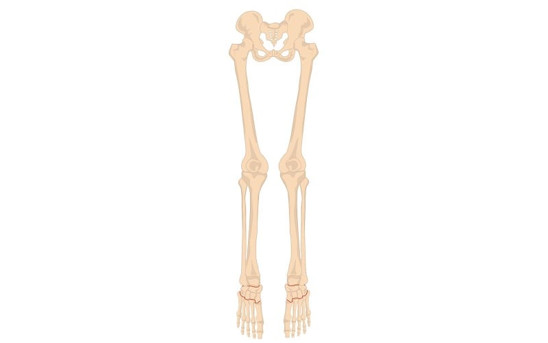


The leg bones are part of the lower limb skeleton and are essential for support, movement, and bearing the body's weight. The main bones in the leg include:
1. Femur (Thigh Bone)
- Location: The femur is the longest and strongest bone in the body, extending from the hip to the knee.
2. Patella (Kneecap)
- Location: A small, triangular bone located in front of the knee joint.
3. Tibia (Shin Bone)
- Location: The larger and stronger of the two lower leg bones, running from the knee to the ankle.
4. Fibula
- Location: A slender bone running parallel to the tibia, on the outer side of the lower leg.
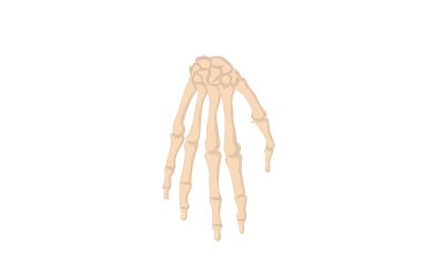
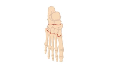
5. Foot Bones (Tarsals, Metatarsals, and Phalanges)
- Tarsals: Seven bones in the hindfoot and midfoot, including the talus (which forms the lower part of the ankle joint) and the calcaneus (heel bone).
- Metatarsals: Five long bones in the midfoot, connecting the tarsals to the toes.
- Phalanges: The bones of the toes, with each toe having three phalanges (proximal, middle, and distal), except for the big toe, which has two.
0 Reviews for this product
0 Comments for this product